How do X rays work?
 X rays are produced whenever high-energy electrons suddenly give up energy ... X-ray machines which produce these rays accelerate electrons towards a metal target. The electrons rapidly slow down when they collide with atoms in the target, and part of their energy is changed into X rays.
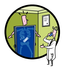
"One of the earliest applications of X rays was in medicine, where they were used for both diagnosis and therapy. Today X rays are still most widely used in this field. They penetrate soft tissues but are stopped by bones, which absorb them. Thus if a photographic plate that is sensitive to X rays is placed behind a part of the body and an X-ray source is placed in front, X-ray exposure will result in a picture of the internal bones and organs. When the plate, or radiograph, is developed, a negative image is produced: bones and dense tissues show up as light or white regions, while tissues that are easily penetrated by X rays appear dark. Although bones are the most opaque structures in the body, there are many dense tissues, such as cancer tumors, that can also show up unusually light in radiographs. Doctors use these images to diagnose diseases, detect foreign objects in the body, examine dental cavities, and study damaged or broken bones.
In order for X rays to be used for the study of other, less dense tissues of the body, such as the gastrointestinal tract, the tissues must first be made opaque to X rays. Generally, doctors ask patients to drink a liquid mixture containing an opaque material, such as barium, so that the internal contours of the alimentary tract become visible with X rays."
Excerpted from Compton's Concise Encyclopedia
Copyright (c) 1995 Compton's NewMedia, Inc.
"George Files" by Parenting the Next Generation

|
X rays are produced whenever high-energy electrons suddenly give up energy ... X-ray machines which produce these rays accelerate electrons towards a metal target. The electrons rapidly slow down when they collide with atoms in the target, and part of their energy is changed into X rays.